Designing drugs around the proteins they must spare
Marc C. Deller, D.Phil.
Presentation Of Drug-discovery Impact, Understanding & Mechanism
Twenty years, one habit: read the structure before the chemistry.
Twenty years from cytokine receptors to kinase drugs.
Structures that became papers, patents and clinical filings.
I built the platform that made every campaign faster.
One pipeline from crystal tray to model.
Structure services other scientists relied on.
A patent deadline, beaten by a crystal structure.
An antibody that recognises only the mutant CALR tail.
A drug is defined by what it must not hit.
Hit FGFR1 and phosphate rises; miss the mutants and cancer returns.
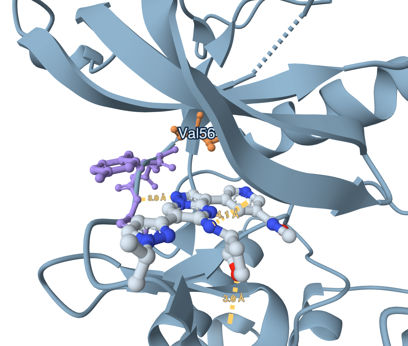
A hydrogen bond drawn on a model, then measured in the crystal.
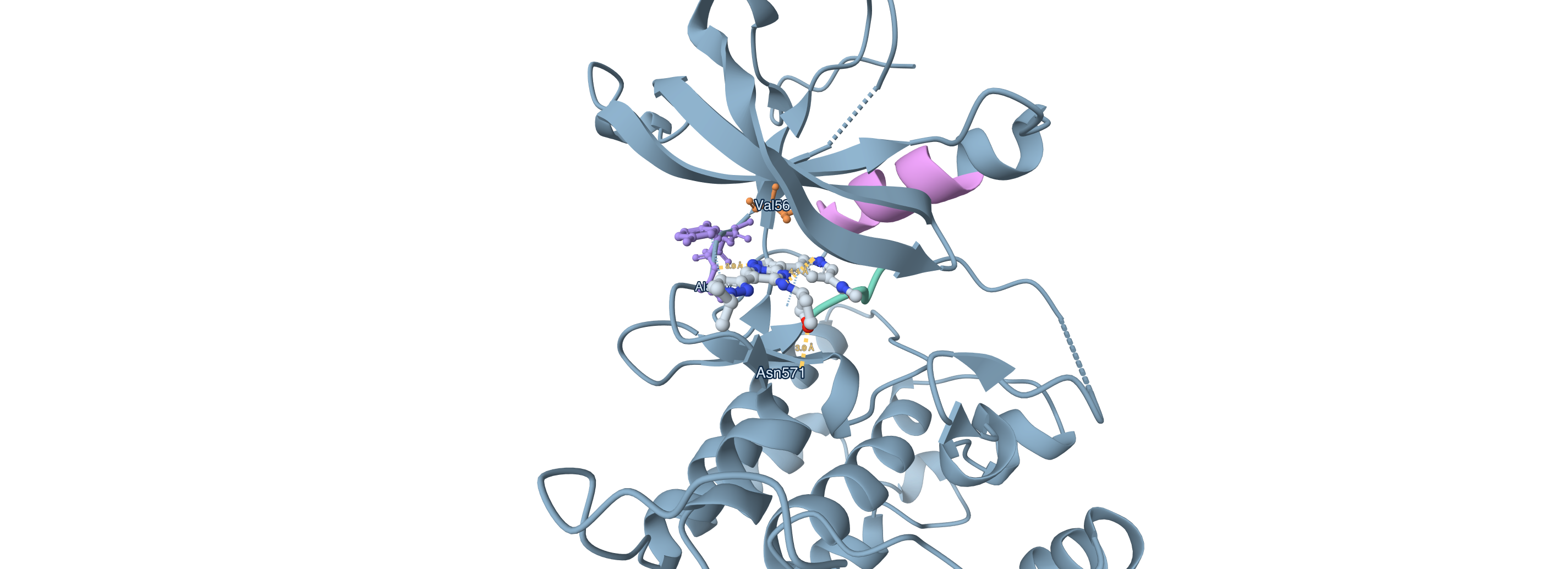
Each edit moved one property without breaking the others.
FGFR1 falls away while the gatekeeper mutants stay inhibited.
FGFR3 vs FGFR1 IC50, every compound (nM, log)
Every polar contact PLIP measured
| PDB | type | residue | Å | role |
|---|---|---|---|---|
| 8E1X | hydrogen bond | Leu487 (bb) | 4.10 | protein_acceptor |
| 8E1X | hydrogen bond | Ala567 (bb) | 3.03 | protein_donor |
| 8E1X | hydrogen bond | Asn571 | 3.01 | protein_donor |
KLIFS (ok, from 8E1X): gatekeeper Val564; hinge Glu565, Tyr566, Ala567; DFG in; αC in.
Three deuterium atoms closed the last metabolic door.
Rat oral bioavailability
The screening hit was a better JAK2 inhibitor than CDK2.
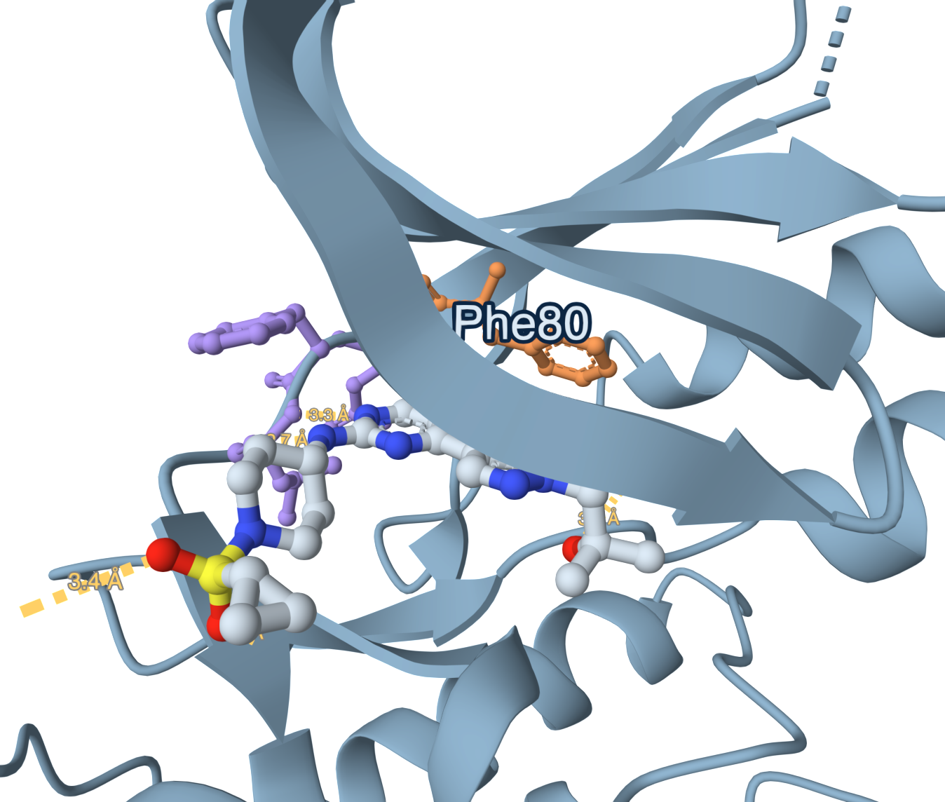
Sulfonamide oxygens reach Asp86; the aminopyrimidine holds the hinge.
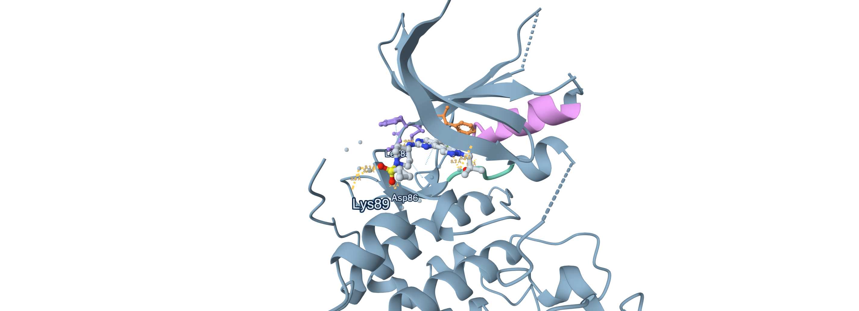
An sp3 swap erased JAK2; everything after was trade-offs.
One trifluoromethyl at the gatekeeper and CDK1 falls away.
CDK2 vs CDK1 IC50, every compound (nM, log)
Every polar contact PLIP measured
| PDB | type | residue | Å | role |
|---|---|---|---|---|
| 8UV0 | hydrogen bond | Leu83 (bb) | 2.73 | protein_acceptor |
| 8UV0 | hydrogen bond | Leu83 (bb) | 3.26 | protein_donor |
| 8UV0 | hydrogen bond | Asp86 (bb) | 3.04 | protein_donor |
| 8UV0 | hydrogen bond | Lys89 | 3.43 | protein_donor |
| 8UV0 | hydrogen bond | Asp145 | 3.66 | protein_acceptor |
| 8UV0 | salt bridge | Asp86 | 4.96 | protein_negative |
| 8UV0 | water bridge | Glu12 | 3.70 | protein_donor |
| 8UV0 | water bridge | Gln85 | 3.06 | protein_donor |
KLIFS (ok, from 8UV0): gatekeeper Phe80; hinge Glu81, Phe82, Leu83; DFG in; αC out.
Potent, isoform-selective, soluble, and exposed by mouth.
Rat oral bioavailability
Inhibit JAK2 and you take the blood counts with you.
Povorcitinib holds the JAK1 hinge with two hydrogen bonds.
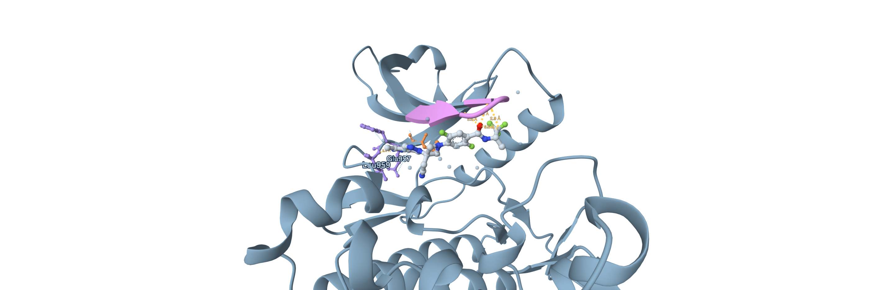
Numbering: JAK2 = JAK1 − 27 at hinge, − 26 at P-loop.
One fluorine folded the amide; a smaller hinge binder fixed the dog.
Same ligand, same hinge, in both isoforms.
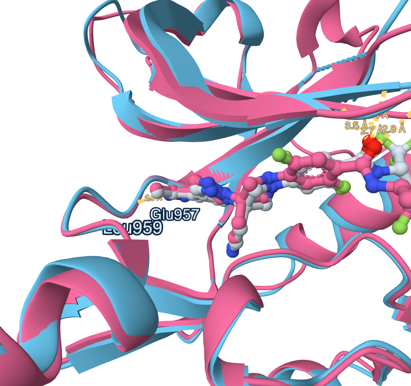
JAK1 vs JAK2 IC50, every compound (nM, log)
Numbering: JAK2 = JAK1 − 27 at hinge, − 26 at P-loop.
Why selective: authors' P-loop hypothesis
Every polar contact PLIP measured
| PDB | type | residue | Å | role |
|---|---|---|---|---|
| 10PI | hydrogen bond | Gly884 (bb) | 3.46 | protein_donor |
| 10PI | hydrogen bond | Gly887 (bb) | 3.44 | protein_donor |
| 10PI | hydrogen bond | Glu957 (bb) | 2.81 | protein_acceptor |
| 10PI | hydrogen bond | Leu959 (bb) | 2.93 | protein_donor |
| 10PI | water bridge | His885 | 3.48 | protein_donor |
| 10PJ | hydrogen bond | Phe860 (bb) | 2.82 | protein_donor |
| 10PJ | hydrogen bond | Gly861 (bb) | 2.71 | protein_donor |
| 10PJ | hydrogen bond | Glu930 (bb) | 2.91 | protein_acceptor |
| 10PJ | hydrogen bond | Leu932 (bb) | 2.96 | protein_donor |
| 10PJ | water bridge | Asp994 | 4.04 | protein_acceptor |
KLIFS (fallback_by_uniprot, from 3EYH (KLIFS structure 1462, same UniProt P23458)): gatekeeper Met956; hinge Glu957, Phe958, Leu959; DFG in; αC in.
Forty-four-fold weaker on the enzyme, fully absorbed in dog.
Dog oral bioavailability
Aspartate is not cysteine: no warhead, so shape must do it.
A rigid cage holds the amine where Tyr96 can reach it.
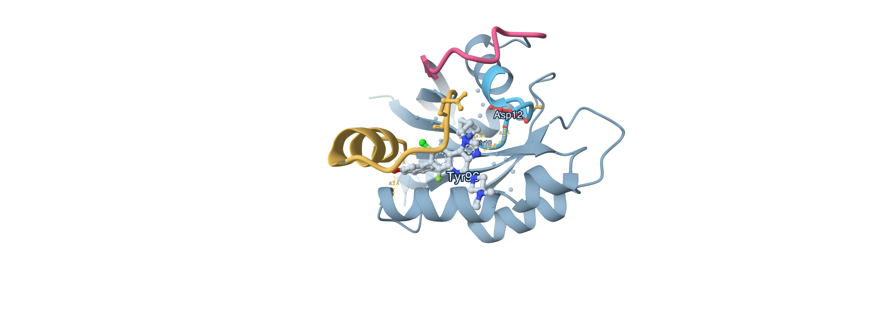
No contact with the Asp12 side chain
It grips the backbone beside Asp12, never the side chain.
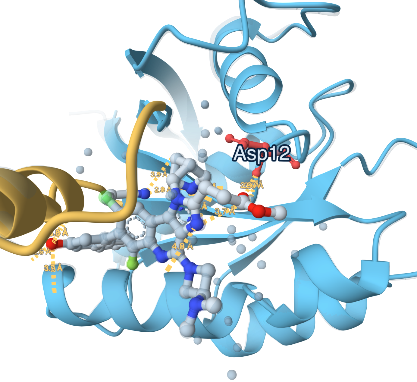
Every polar contact PLIP measured
| PDB | type | residue | Å | role |
|---|---|---|---|---|
| 9E5F | hydrogen bond | Gly10 (bb) | 2.96 | protein_acceptor |
| 9E5F | hydrogen bond | Glu63 (bb) | 3.82 | protein_acceptor |
| 9E5F | hydrogen bond | Tyr96 | 2.77 | protein_donor |
| 9E5F | hydrogen bond | Arg102 | 3.69 | protein_donor |
| 9E5F | salt bridge | Glu62 | 2.87 | protein_negative |
| 9E5F | water bridge | Gly10 | 2.85 | protein_donor |
| 9E5F | water bridge | Asp12 | 3.72 | protein_donor |
| 9E5F | hydrogen bond | Gly10 (bb) | 2.96 | protein_acceptor |
| 9E5F | hydrogen bond | Glu63 (bb) | 3.82 | protein_acceptor |
| 9E5F | hydrogen bond | Tyr96 | 2.77 | protein_donor |
| 9E5F | hydrogen bond | Arg102 | 3.69 | protein_donor |
| 9E5F | salt bridge | Glu62 | 2.87 | protein_negative |
| 9E5F | water bridge | Gly10 | 2.85 | protein_donor |
| 9E5F | water bridge | Asp12 | 3.72 | protein_donor |
KRAS motifs from UniProt P01116 and Pfam PF00071: P-loop 10-17, switch I 30-38, switch II 60-76, G12D, Gly13, Gln61.
SPR discrepancy, compound 23: graphical abstract 2.2 nM, Table 3 22 nM. Compound 10 whole-blood pERK: graphical abstract 451 nM, Table 1 410 nM. Table values shown throughout; neither averaged.
A thousand-fold weaker, and finally absorbed by mouth.
Monkey oral bioavailability
Table values shown; graphical abstract prints 2.2 nM (T3: 22) and 451 nM (T1: 410).